Gynecomastia vs Chest Fat: How to Tell the Difference
A fuller chest does not tell the whole story.Understand the tissue before choosing an approach.
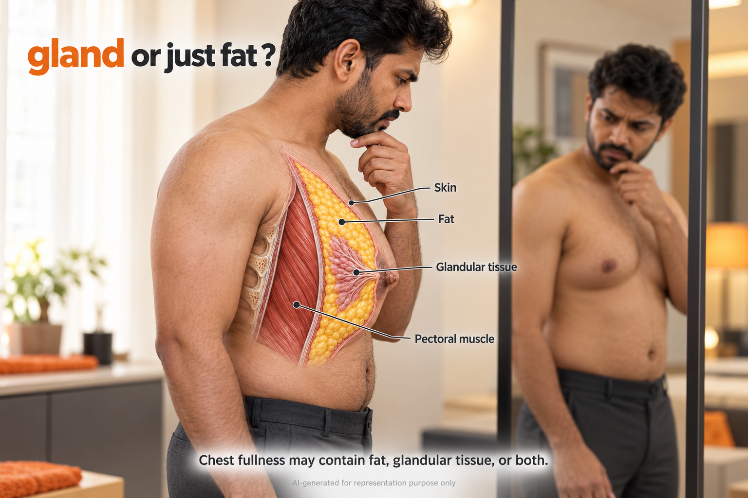
Gynecomastia vs chest fat is a difference in tissue, not simply in appearance. True gynecomastia involves enlarged breast gland; pseudogynecomastia means fullness from fat without glandular enlargement. Both can coexist. A photograph, body weight or home pinch test cannot reliably establish which is present. We use your history and a physical examination to understand the finding and decide whether further tests are needed. [1] [3]
“
Your chest can contain different mixtures of gland and fat. Understanding that difference comes before choosing treatment.
”
– Dr Gaurav Patodia
Why two similar- looking chests need different approaches?
You might notice fullness beneath a T-shirt, a change after weight loss or an uneven chest contour. However, appearance alone does not reveal what lies beneath the skin. More than one tissue can contribute to the same outline.
The useful first question is therefore not “Which procedure do I need?” It is “What is making my chest look or feel different?” Bring us the timeline, your concerns and any previous reports. An assessment can clarify the next step without committing you to treatment.
Clues can guide a conversation. They cannot make the diagnosis.
Localised fullness beneath the nipple may suggest glandular tissue, whereas a softer, more diffuse fullness may reflect fat. Yet these patterns overlap, and tenderness is not a reliable way to separate them. A firm area is not automatically gynecomastia. [3] [2]
Tell us when the change began, whether your weight has changed and which medicines, supplements or hormone products you use. These details give the examination context. Our guide to possible causes and age-of-onset guide explain related questions without turning a clue into a diagnosis.
UNDERSTANDING THE TOPIC
Gynecomastia vs chest fat: what examination can clarify
Gynecomastia is benign growth of male breast glandular tissue. Pseudogynecomastia is fat accumulation without that glandular growth. Gland and fat can coexist, so being lean or overweight does not establish the diagnosis. [1] [3]
Distribution and texture are clues
Gland may form a firmer area beneath the nipple; fat often feels more diffuse. However, a distinct disc is not present in every case. Tenderness and shape overlap. Neither a pinch test nor a photograph can confirm the tissue or exclude cancer. [2] [3]
History, examination and selected tests
We assess onset, symptoms, weight change, medicines and relevant health history. We examine the breast, nipples and nearby lymph nodes, and look for other findings when appropriate. Blood tests address suspected underlying causes; everyone does not need the same panel. [1] Breast imaging is not routinely needed when examination is typical of gynecomastia or pseudogynecomastia. Uncertain or suspicious findings may need ultrasound, mammography or tissue sampling. [4]
When to seek prompt assessment
Arrange prompt examination for rapid change, a hard or fixed lump away from the nipple centre, bloody nipple discharge, skin dimpling, nipple retraction or enlarged underarm nodes. These findings need assessment rather than a home test. [2] [4]
Identifying the tissue guides care. Weight reduction may improve fatty fullness, but exercise does not selectively remove established gland. Observation or treatment depends on the cause, duration, symptoms and examination. [1] [2]
Medical references
[1] Kanakis GA et al. EAA clinical practice guidelines—gynecomastia evaluation and management. Andrology. 2019;7:778–793.
[2] Swerdloff RS, Ng JCM. Gynecomastia: Etiology, Diagnosis, and Treatment. Endotext. Updated 6 January 2023.
[3] Johnson RE, Murad MH. Gynecomastia: Pathophysiology, Evaluation, and Management. Mayo Clinic Proceedings. 2009;84:1010–1015.
[4] Expert Panel on Breast Imaging; Niell BL et al. ACR Appropriateness Criteria® Evaluation of the Symptomatic Male Breast. Journal of the American College of Radiology. 2018;15:S313–S320.
What a proper assessment can help you understand
Separate an observation from a diagnosis
Describe what you have noticed without needing to name the tissue yourself. Chest shape alone cannot settle the question.
Recognise that mixed tissue is possible
You do not have to fit a gland-only or fat-only category. Ask us what contributes to your own chest contour.
Make your history useful
Bring a medication list, previous reports and a simple timeline. Do not stop prescribed medicines without speaking to the treating prescriber.
Understand why a test is suggested
Ask which finding a blood test or scan is intended to clarify. More testing does not automatically mean a better assessment.
Set realistic expectations
Our guide to improvement without surgery explains why changes in fat and changes in gland are different outcomes.
Keep treatment proportionate
A diagnosis does not automatically mean surgery. Our gynecomastia treatment service explains how we discuss suitable care after assessment.
Gynecomastia vs chest fat: three common questions
Can I diagnose gynecomastia with a pinch test?
No. Feeling a firm area may raise a question, but it does not confirm glandular gynecomastia or exclude another breast condition. Texture varies, and fat and gland may overlap. [2] [3]
Avoid repeated squeezing or judging yourself against online photographs. If you are uncertain, we can assess the change and explain the findings in person.
Can I have both chest fat and gynecomastia?
Yes. Breast gland and surrounding fat can both contribute to fullness. A change in body weight may alter the fat component while gland remains. [2] [3]
Likewise, a lean appearance does not prove that every area of fullness is gland. We consider the whole examination instead of classifying the chest from body size alone.
Do I need an ultrasound to tell gland from fat?
Not always. If examination findings are typical of gynecomastia or pseudogynecomastia, ACR guidance does not recommend routine breast imaging. [4]
When the findings are unclear or suspicious, imaging or a biopsy may be appropriate. The choice depends on the examination and age; ultrasound is not the only possible investigation. [4]
YOUR NEXT STEP
Understand the finding before deciding what to do.
If you are unsure whether chest fullness is gland, fat or both, arrange an assessment at Dermacure Clinics, Bank More, Dhanbad. We can explain the findings and appropriate next steps. You can contact the clinic to discuss an appointment.
Ask About a Personal Assessment